
Mitotic figure countingin breast H&E
Counting mitoses by hand is slow and varies between readers. Aiosyn Mitosis Breast uses artificial intelligence to detect mitotic figures across the whole-slide image before the case is opened.
Aiosyn Mitosis Breast is for research use only.
- Detection across the whole-slide image, not a sampled field
- Validated in a multi-center clinical study
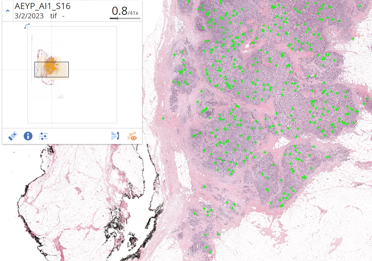
Every mitosis, found before the case is opened
Drag the handle across a single H&E field. The mitotic figures a pathologist has to search for are already marked, so the work becomes faster and more reproducible.
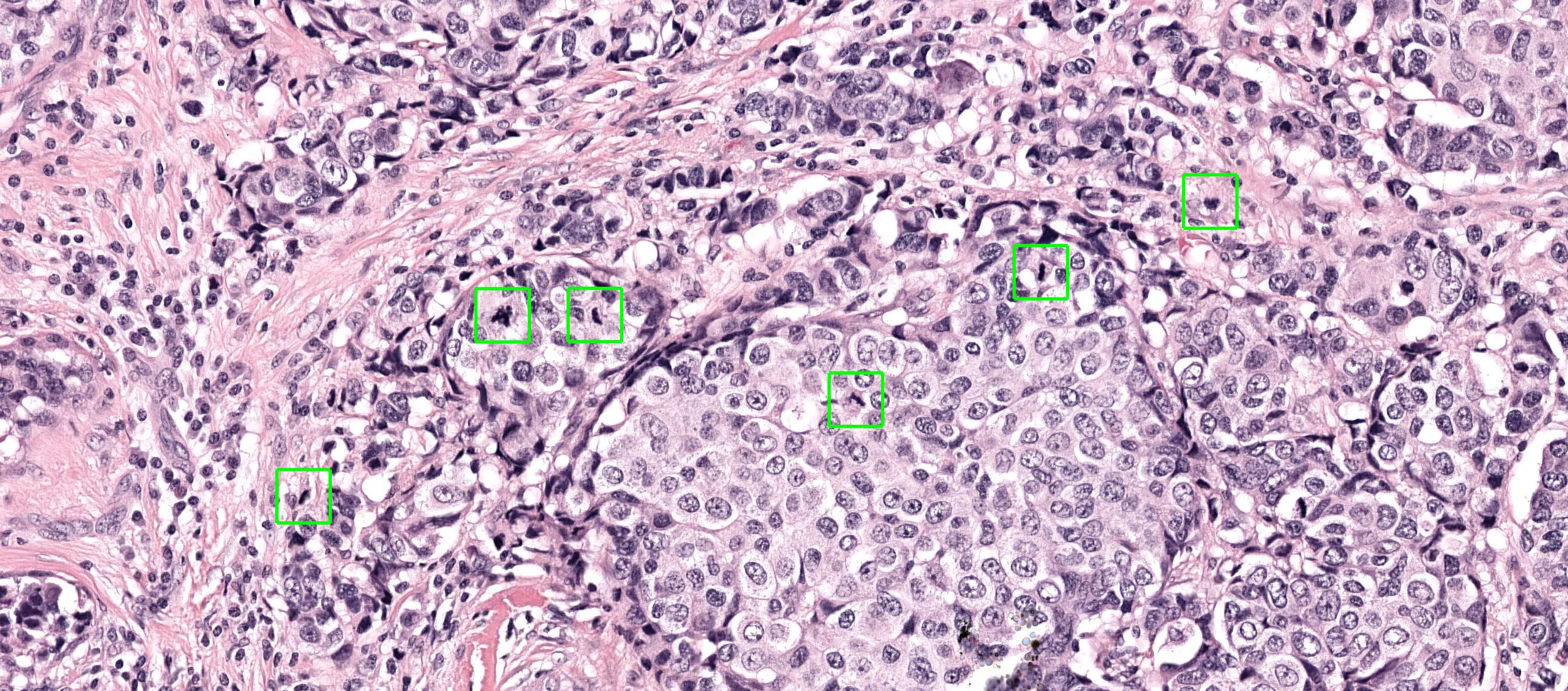
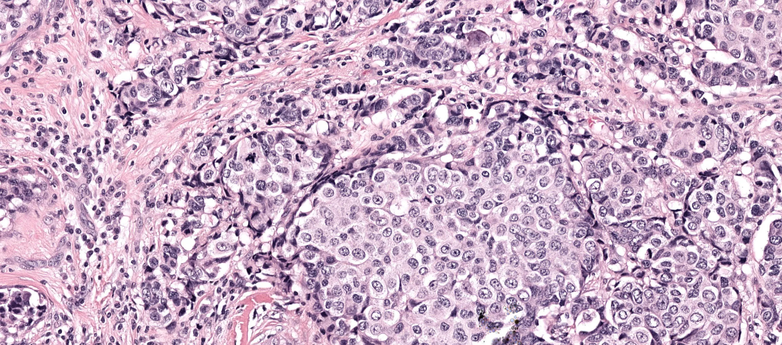 H&E sectionDetected mitoses
Consistency, and time back on every case
Mitotic counting is slow, subjective work that breast grading depends on. With Aiosyn Mitosis Breast, pathologists are faster and agree more with each other.
Less time on the slowest step
Reviewing detections replaces searching for them. Pathologists spend their time confirming a count rather than hunting across fields for the figures to count.
More consistent grading
Mitotic count is one of the most variable parts of breast grading. Detection is reproducible, so every reader starts from the same set of candidates.
The whole section, every slide
Detection runs across every field of the whole-slide image, so a long series is read the same way from the first slide to the last.


Measured with 28 pathologists
Aiosyn Mitosis Breast was validated in an independent multi-center clinical study run at Radboud university medical center, with certified pathologists reading breast biopsies and resections both with and without algorithm support.
Aiosyn Mitosis Breast
Mitotic figure count, breast H&E
- Time savings
- +60%
- Reading resections faster
- Productivity
- +15%
- Average across readers
- Inter-observer consistency
- +32%
- Between pathologists
Over 90% of surveyed participants said they would use the algorithm in daily practice. The efficiency gain did not come at the cost of accuracy.
How the analysis runs
Slides in, marked detections back.

1
Sections are prepared and scanned as usual.
2
The algorithm detects mitotic figures across the whole-slide image.
3
Every detected figure comes back marked on the slide.
4
The pathologist confirms the count, assisted by Aiosyn Mitosis Breast.
Working in a different tissue, stain or endpoint? Custom AI development tailors the pipeline to your study.
Mitosis in the news
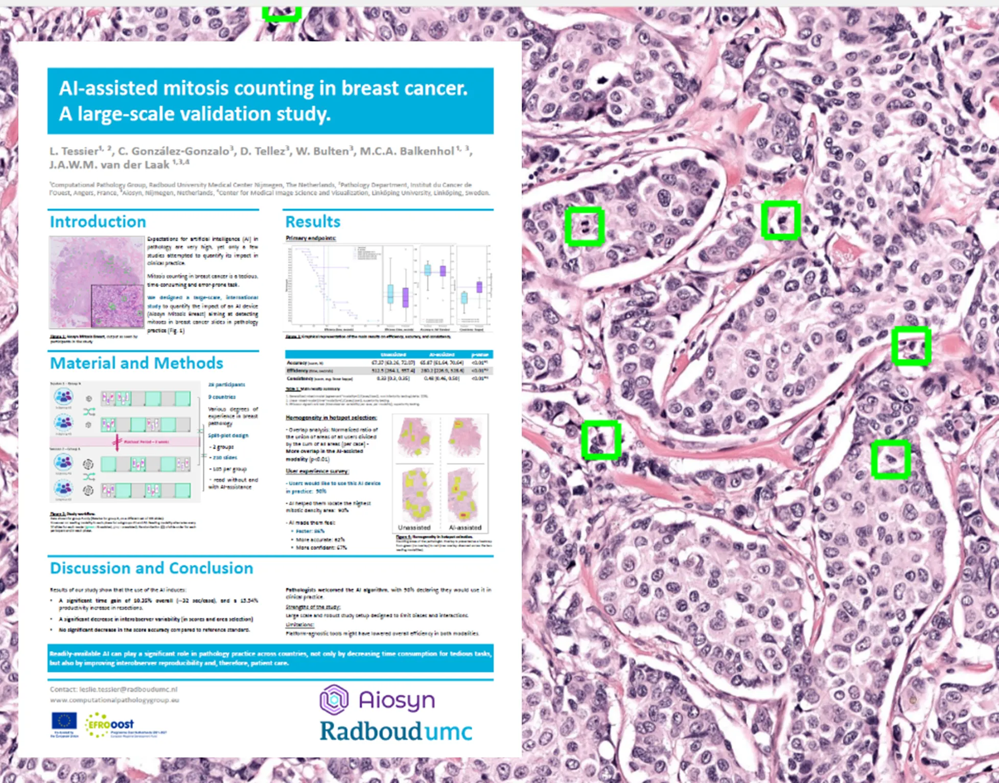


Aiosyn Mitosis Breast is for research use only.
Also in the clinical track
Clinical overview- AiosynQCAutomated whole-slide quality control
- Mitosis (breast)Mitotic figure counting for breast cancer
- Custom AI DevelopmentA model built for your tissue, stain or endpoint
See it on your own slides
Upload a handful of whole-slide images and we will show you the detections in our viewer, alongside your own images.