

Skip to content
Aiosyn

AI histopathology for preclinical research

The new standardin pre-clinical pathology

Objective, reproducible AI-based tissue quantification across your full cohort, for better insights, more statistical power, and faster study completion.

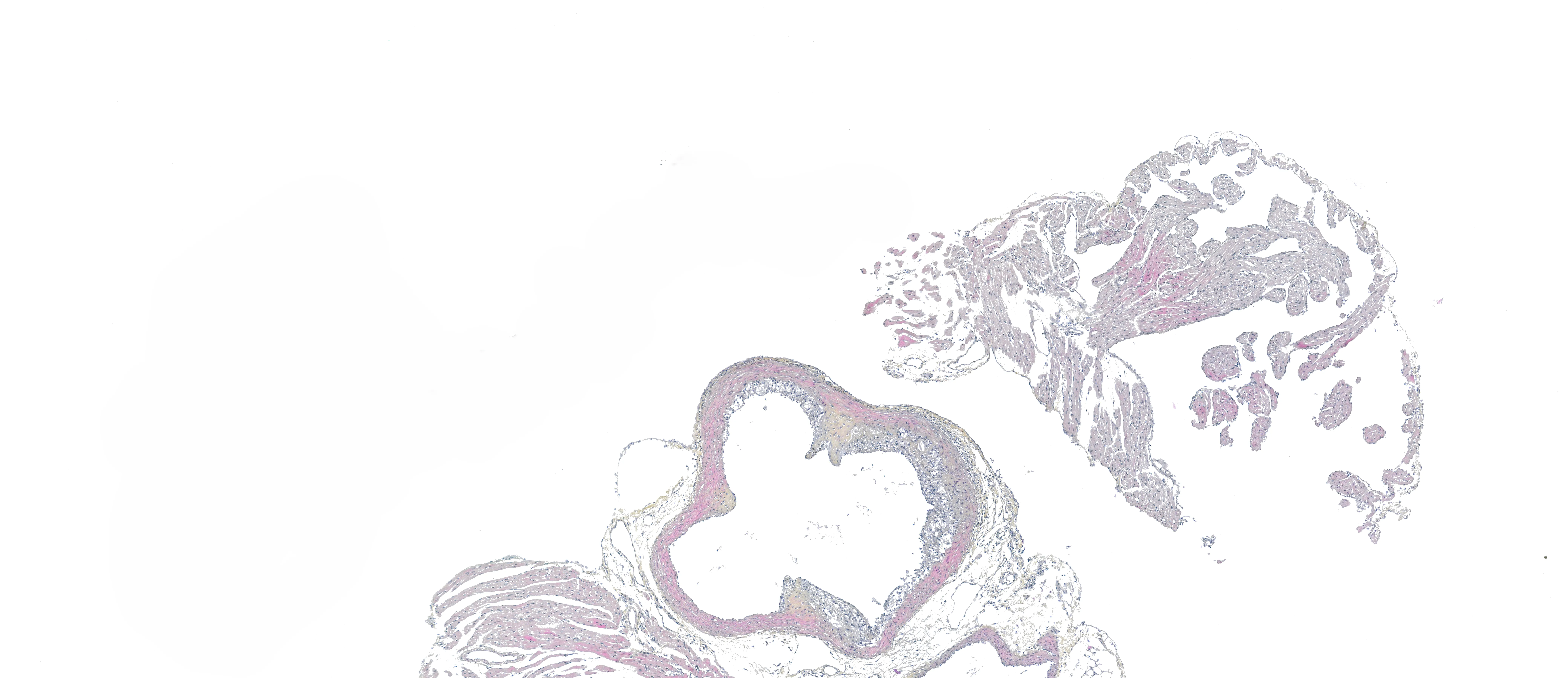
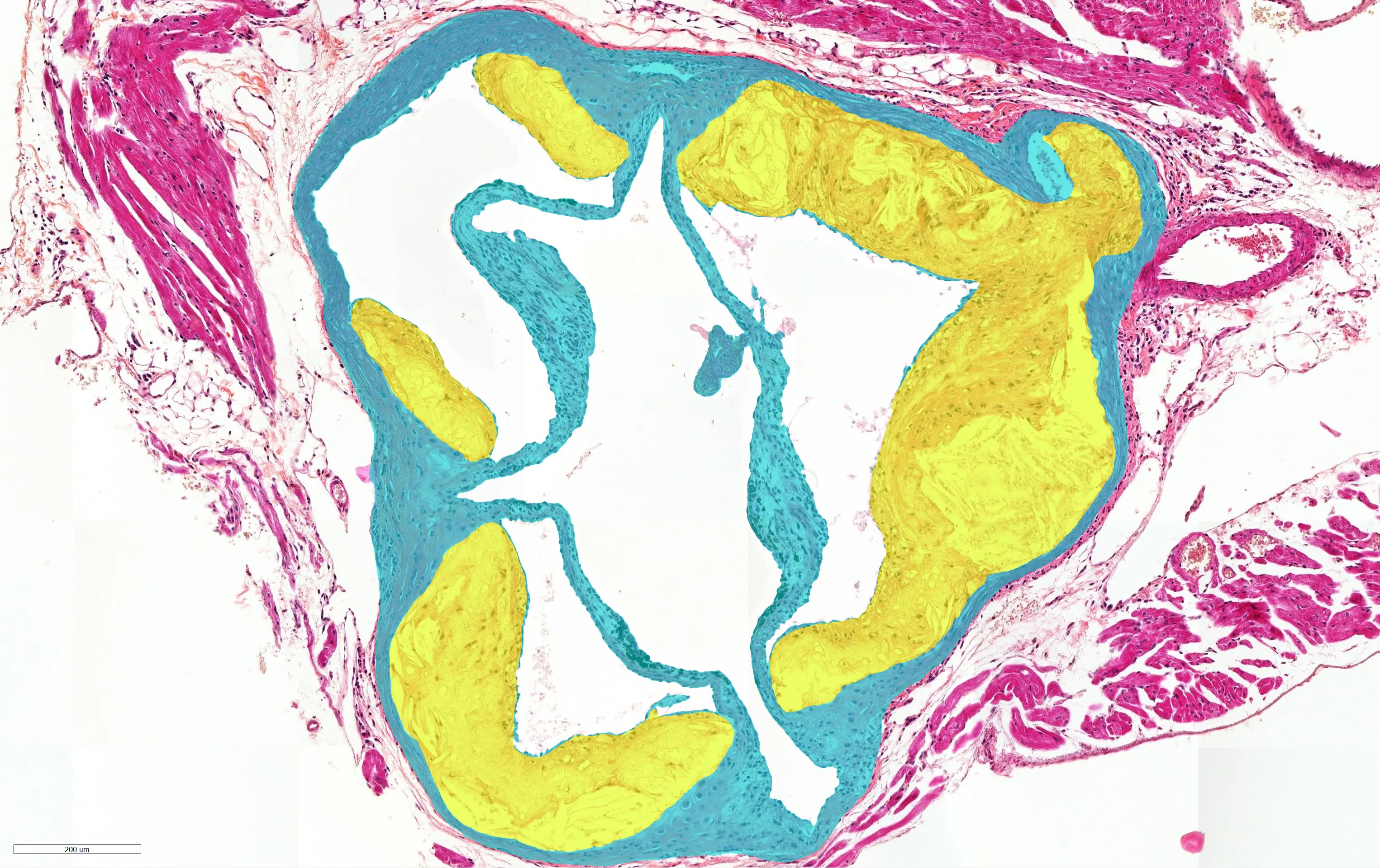
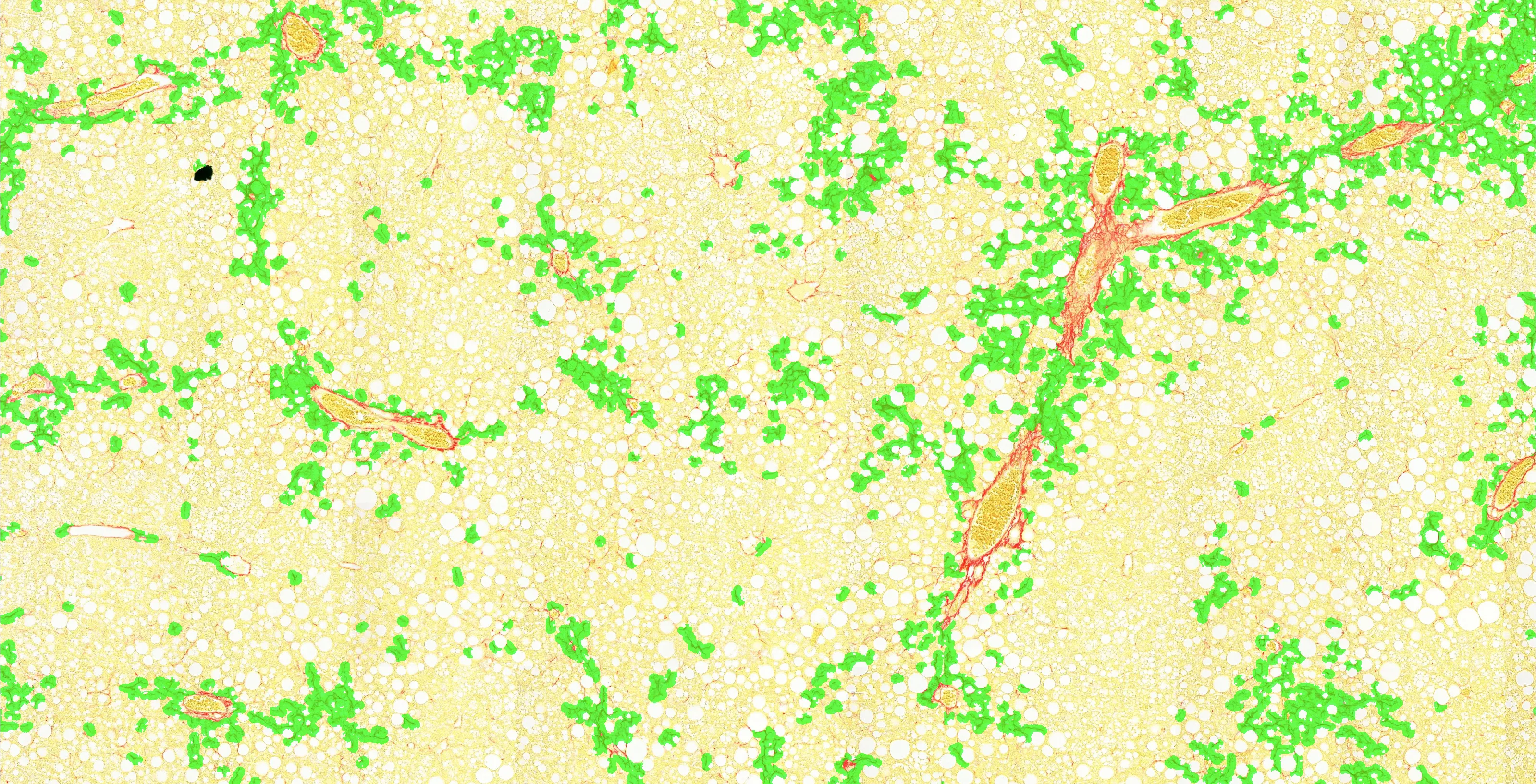
Atherosclerotic plaque shown as stained tissue on the left and the AI segmentation read-out on the right. Use the slider below to reveal either side.
AI read-outTissue

Built for your disease area

Working models across four areas, each built and validated on real preclinical cohorts. Different area? Custom analysis tailors the pipeline to your study.

Custom analysis

Working in a different tissue, stain, or endpoint? We tailor the pipeline to your study and build the read-out you need.

Talk it through

The evidence

Validated results with our partners

We co-develop and validate our AI with expert pathologists and preclinical CROs, so the read-outs are objective, reproducible, and applicable to real preclinical cohorts.

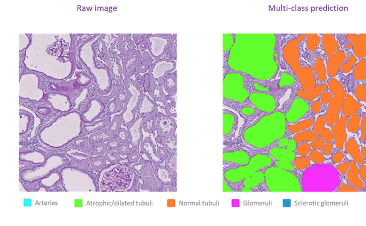
Figure from the published study: a raw PAS-stained kidney section beside Aiosyn's multi-class AI prediction, with a legend for arteries, tubuli, and glomeruli.

Peer-reviewed study

AI read-outs across whole kidney sections detected a treatment effect that manual scoring missed

In a peer-reviewed diabetic-nephropathy study with Physiogenex, the AI counted every glomerulus and tubule across the whole kidney section. Its read-outs resolved a dapagliflozin treatment effect that classical manual scoring could not detect.

Physiogenex

Briand F, et al. · European Journal of Pharmaceutical Sciences · 2026

Read the study
AI versus pathologist lesion area Scatter of 80 cases comparing Aiosyn AI lesion-area read-outs with expert pathologist scoring; points cluster along the line of perfect agreement (Pearson r = 0.96). 0 0 400 400 800 800 1200 1200 1600 1600 y = x r = 0.96 · R² = 0.91 · n = 80 Pathologist lesion area (×10³ µm²) AI lesion area (×10³ µm²)

Method validation

Across 80 cases, AI lesion-area read-outs matched expert pathologist scoring

Developed with TNO, the AI measures atherosclerotic lesion area across the full aortic-root cross-section. Its read-outs tracked expert pathologist scoring closely, giving an objective, reproducible alternative to manual measurement.

Atherosclerosis · aortic-root cross-sections

Explore atherosclerosis read-outs

Let's talk about your study

Tell us about your cohort and endpoints. We will show you what Aiosyn's read-outs would look like on your tissue.

Discuss your study

Power, not just speed

Manual scoring is slow, varies between readers, and only ever samples the slide. The value here is precision: reproducible, whole-tissue read-outs give your study more statistical power, so real treatment effects show up more clearly and leaner cohorts can carry the work.

How it works
  • Reproducible by construction

    The same slide returns the same read-out every time. No inter-reader variability, so a real treatment effect is not lost in measurement noise.

  • Across 100% of the tissue

    Every structure on the whole section is quantified, not a pathologist-sampled subset. More signal means more statistical power, and fewer animals needed to reach it.

  • Fast enough to scale

    Whole cohorts are analysed in a fraction of the time of manual review, so a large study is no longer gated on reader hours.

A whole section, fully analysed

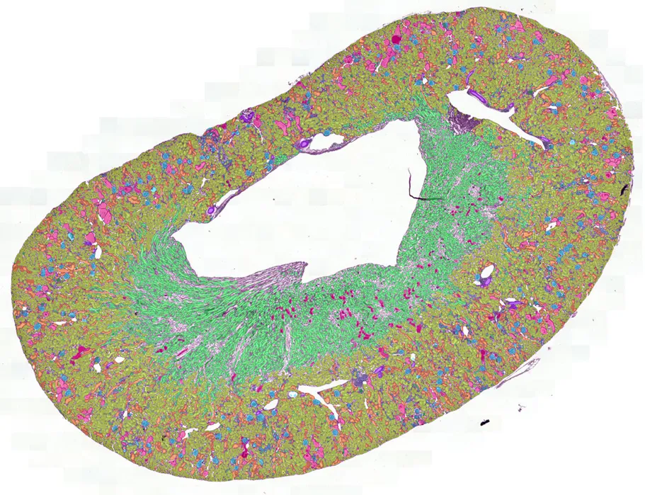
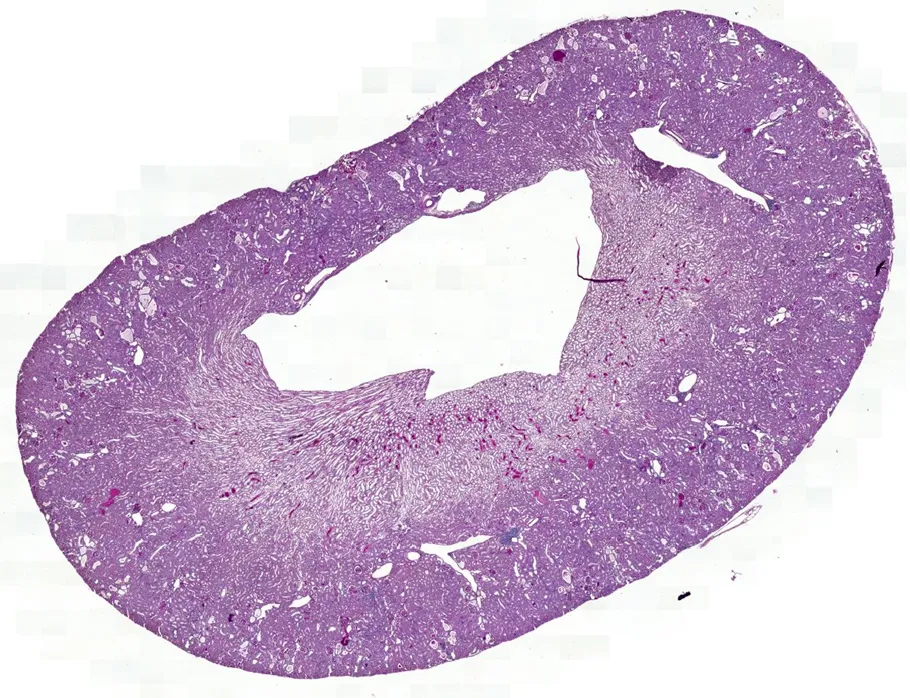
Drag the handle across an entire kidney section: every structure classified, every cell detected, edge to edge. What a pathologist scores by sampling a handful of fields, the AI reads across 100% of the tissue.

Stained sectionAI analysis
A complete kidney cross-section. Left: the stained tissue. Right: Aiosyn's AI, segmenting and classifying every structure across the whole section, not a sampled subset.

From slide to read-out

Three steps, no new scanning workflow, no manual scoring in between.

  1. 1

    Share your slides

    Send whole-slide images of your stained tissue in the formats you already scan.

  2. 2

    The AI does the reading

    Aiosyn's AI segments structures, detects cells, and quantifies across every slide in the cohort.

  3. 3

    Get objective read-outs

    Receive reproducible, whole-tissue quantification, ready for your analysis and defensible in review.